Cancer Care
Visitor Guidelines
For the safety and comfort of our patients, we ask that children 12 and under not accompany you to clinic, infusion or radiation oncology visits. We understand this may be inconvenient and are here to help. Don't hesitate to get in touch with your healthcare team if you have any questions or need support.
No two cancer cases are exactly the same. And there’s one place that truly understands that.
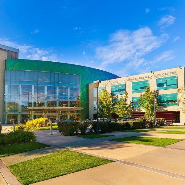
Moores Cancer Center at UC San Diego Health is San Diego's only National Cancer Institute (NCI)-designated Comprehensive Cancer Center. We are repeatedly ranked among the top 50 in the nation for cancer care by U.S. News & World Report.
Let's Get Started
The oncologists at UC San Diego Health not only provide personalized care with better outcomes, they set treatment standards nationwide for more than 200 types of cancer — including, most importantly, yours.
Cancers We Treat
At UC San Diego Health, you'll get the best care and the most advanced treatment options in a warm and supportive environment.
Video: Cancer Care as Unique as You

Get trusted care for cancer at UC San Diego Health where world-class specialists use the latest innovations and advanced technology to personalize treatments for your kind of cancer.
Clinical Trials
Advancing Cancer Care and Prevention
UC San Diego Health physicians are actively researching ways to improve cancer care. By joining a clinical trial, you may receive a new cancer treatment before it is available to the public. If your cancer has not responded to standard therapies, talk to your doctor about whether a clinical trial could be right for you.
Cancer Treatment and Support for the Whole Person
A cancer diagnosis can affect you in body, mind and spirit. You have a spectrum of supportive services available to you and your loved ones.
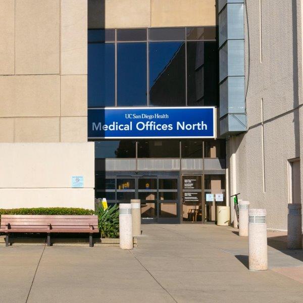
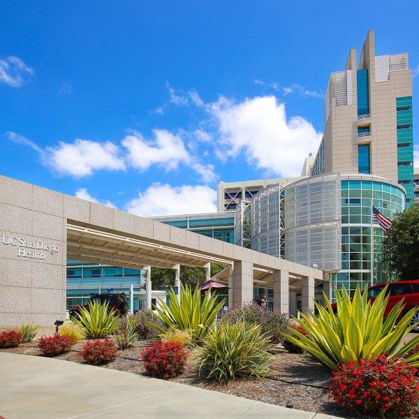
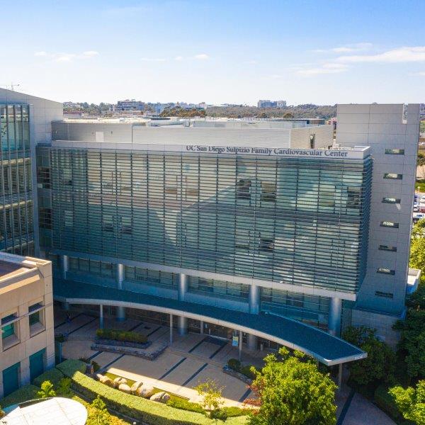
Locations
For your convenience, we offer cancer care services at locations throughout San Diego County. No matter where your appointment is, your care is coordinated by an expert team, and you have access to comprehensive support services at Moores Cancer Center in La Jolla.


Study: Breast Cancer Drug Shows Potential for Rare Appendix Cancer
Researchers at University of California San Diego School of Medicine found an FDA-approved drug used...

UC San Diego Health No. 1 in San Diego, Top 20 in the Nation
UC San Diego Health has once again secured the No. 1 ranking in San Diego, according to the 2024-202...

UC San Diego Health Offers Novel Gene Therapy for Bladder Cancer
UC San Diego Health is the first health system in San Diego County to offer a new bladder-saving gen...